
Life: Magnified is an exhibit of scientific images showing cells and other scenes of life magnified by as much as 50,000 times. The exhibit is on display at Washington Dulles International Airport’s Gateway Gallery from June through November 2014. A Web companion is available through NIH here http://www.nigms.nih.gov/education/life-magnified/Pages/default.aspx
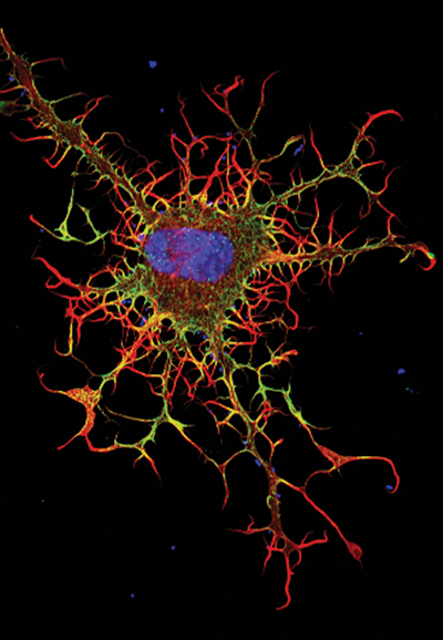
When biologists disabled proteins critical for cell movement, the result was dramatic. The membrane, normally a smooth surface enveloping the cell, erupted in spiky projections. This image (see Fig. 1), which is part of the Life: Magnified exhibit, resembles a supernova. Although it looks like it exploded, the cell pictured is still alive.
To create the image, Rong Li, PhD, and Praveen Suraneni, PhD, NIH-funded cell biologists at the Stowers Institute for Medical Research in Kansas City, Missouri, disrupted two proteins essential to movement in fibroblasts—connective tissue cells that are also important for healing wounds. The first, called ARPC3, is a protein in the Arp2/3 complex. Without it, the cell moves more slowly and randomly.1 Inhibiting the second protein gave this cell its spiky appearance. Called myosin IIA (green in the image), it’s like the cell’s muscle, and it’s critical for movement. The blue color is DNA; the red represents a protein called F-actin.
Drs. Li and Suraneni are studying cell movement because it is vital throughout development. In embryos, cells wriggle to their correct location. In the developing brain, neurons migrate to settle in their designated brain structure. Immune cells move toward bacterial and viral invaders—and skin fibroblasts squirm toward injuries to patch wounds.
Disrupting the proteins crucial for movement can wreak havoc: immune diseases, birth deformities, and even death. On the other hand, blocking cell movement could actually be beneficial in certain circumstances. For example, when a cancer cell breaks away from its primary tumor and migrates, resettling in another part of the body—called metastasis—the results can be deadly. Finding a target on these cancer cells to hinder movement could slow, or even block, metastasis.
The next challenge for Dr. Li’s team is to explore the role of Arp2/3 in other cell types, such as stem cells, to determine whether this complex of proteins also holds the key to their ability to move. ■
Life: Magnified is a joint project among the National Institute of General Medical Sciences, the American Society for Cell Biology and the Metropolitan Washington Airports Authority’s Arts Program, which utilizes the arts to enhance travel experiences at Dulles International and Reagan National Airports. ZEISS provided additional support of the exhibit.
Fig. 1 depicts a fibroblast, a connective tissue cell that plays an important role in wound healing. Normal fibroblasts have smooth edges. In contrast, this spiky cell is missing a protein that is necessary
for proper construction of the cell’s skeleton. Its jagged shape makes it impossible for the cell
to move normally. In addition to compromising wound healing, abnormal cell movement can lead
to birth defects, faulty immune function and other health problems.
Reference
1. Suraneni P, Rubinstein B, et al: The Arp2/3 complex is required for lamellipodia extension and directional fibroblast cell migration. J Cell Biol 16:197:239-251, 2012.