
Dermatologic Events in Oncology is guest edited by Mario E. Lacouture, MD, an Associate Member in the Division of Dermatology, Department of Medicine, at Memorial Sloan-Kettering Cancer Center, New York. He is a board-certified dermatologist with a special interest in dermatologic conditions that result from cancer treatments.
Acute graft-vs-host disease is the major cause of nonrelapse mortality following hematopoietic stem cell transplantation for patients with hematologic malignancies. Acute graft-vs-host disease results from epithelial tissue damage following conditioning chemotherapy with subsequent infusion of competent donor lymphocytes within the allograft that result in damage to the skin, gut, and liver.
The incidence of acute graft-vs-host disease is estimated to be 40% to 60% for those undergoing allogeneic stem cell transplantation, and cutaneous involvement is frequently the earliest manifestation. Acute graft-vs-host disease may occur either in isolation or in combination with intestinal or hepatic disease. Skin biopsy may demonstrate interface dermatitis with necrotic keratinocytes, but its utility in diagnosing acute graft-vs-host disease is controversial.
Risk factors for acute graft-vs-host disease include histocompatibility antigen mismatch, older age of transplant recipient, total-body irradiation as a component of conditioning chemotherapy, gender mismatch between donor and recipient, and prior host exposure to blood products.
Acute graft-vs-host disease usually presents within the first 100 days following hematopoietic stem cell transplantation. However, in this era of reduced-intensity stem cell transplantation and the dynamic nature of immunosuppressive medications used in the prophylaxis against and treatment of graft-vs-host disease, other forms of acute graft-vs-host disease are now recognized; these include late-onset acute, recurrent acute, and persistent acute graft-vs-host disease.
Clinical grading of acute cutaneous graft-vs-host disease is based on stage I (rash on < 25% of body surface area), stage II (rash on 25%–50% of body surface area), stage III (rash on > 50% of body surface area) and stage IV (erythroderma with bullae or desquamation). Notable omissions from these criteria include the morphology of skin lesions, anatomic distribution of lesions, and the symptoms produced by the eruption.
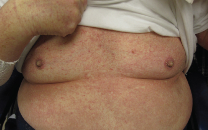
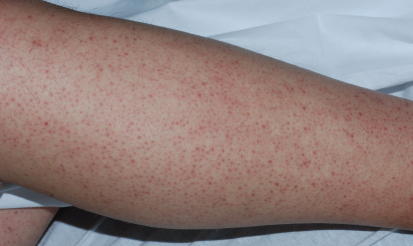
Erythema of the ears or palms may herald the onset of acute graft-vs-host disease. While a morbilliform appearance (Fig. 1) is the most common skin manifestation of acute graft-vs-host disease, other morphologies are well recognized and include violaceous lesions, reticulated variants, and severe forms that produce widespread desquamation that may mimic toxic epidermal necrolysis. Follicular accentuation (Fig. 2) of skin lesions may be an early but important clinical clue during the development of acute graft-vs-host disease.
Treatment Recommendations
The cornerstone of treatment for acute graft-vs-host disease is the prophylactic regimen, but once cutaneous lesions develop, treatment is based on the stage of disease.
Stage I disease can be treated with mid- to high-potency topical steroids alone (eg, alclometasone cream to the face, or fluocinonide or clobetasol creams or ointments to the body) or calcineurin inhibitors (pimecrolimus [Elidel] cream or tacrolimus [Protopic] ointment).
Stages II to IV require both topical and systemic steroids, with systemic doses ranging from 1 to 2 mg/kg daily given over the course of several weeks and slowly tapered depending on patient response.
Ensuring that the calcineurin inhibitor used for graft-vs-host disease prophylaxis is within the serum therapeutic range, or restarting a systemic calcineurin inhibitor along with high-dose systemic steroids at the onset of stage II to IV acute graft-vs-host disease is preferred.
The application of topical steroids twice daily to moist skin and covered with warm wet towels as an occlusive measure (“wet wrap”) is particularly effective for patients with diffuse skin graft-vs-host disease. The use of systemic antihistamines (eg, hydroxyzine) and nonprescription antipruritic topical agents containing camphor or menthol are also efficacious for symptom control.
Narrow-band UVB phototherapy is an additional adjunct that may help expedite the tapering of systemic steroids. Extracorporeal photopheresis, an effective treatment for patients with chronic graft-vs-host disease, has recently been shown to benefit patients with steroid-refractory graft-vs-host disease. ■
Dr. Cotliar is in the Departments of Dermatology & Medicine at Northwestern University Feinberg School of Medicine in Chicago.
Disclosure: Dr. Cotliar reported no potential conflicts of interest.