


Intertriginous areas refer to skin folds (such as axillae, inguinal creases, and inframammary creases), which are characterized by increased friction, temperature, and occlusion. Intertriginous drug reactions are an underrecognized side effect associated with pegylated liposomal doxorubicin therapy, occurring in approximately 3% to 12% of patients who receive such treatment.
Onset of the eruption can occur anywhere from 1 to 25 days after treatment. No predilection exists for sex, age, underlying malignancy, or preexisting skin condition.
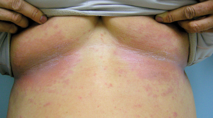
Clinical Manifestation
The rash typically consists of red papules as the primary lesion, later developing into confluent beefy red or violaceous patches that are primarily centered in the axillary and inguinal folds. Areas that are less commonly affected include infra-
mammary creases (see Fig. 1), abdominal folds, back of the neck, antecubital fossa, and other areas under occlusion.
In most cases, the rash progresses to mild superficial skin sloughing and eventual spontaneous resolution with postinflammatory hyper- or hypopigmentation. The eruption is typically mildly pruritic, but it occasionally can be tender or even develop painful erosive lesions.
The exact mechanism is unproven, but the leading hypothesis on why pegylated liposomal doxorubicin is the chemotherapy agent most associated with intertriginous eruptions involves its long half-life and hydrophilic coating. This coating decreases cardiotoxicity but may increase its ability to concentrate within skin, particularly in eccrine sweat glands.
The association of liposomal doxorubicin with palmar-plantar erythrodysesthesia, or hand-foot syndrome, has been well documented in the literature and was described by Beth McLellan, MD, in the June 25, 2013, issue of The ASCO Post. In both conditions—hand-foot syndrome and intertriginous eruptions—local microtrauma and vasodilation may promote drug extravasation, leading to increased drug concentration.
While intertriginous eruptions may share a similar pathogenesis with hand-foot syndrome, it is less common, and thus may be less recognized by oncologists. It should be noted that intertriginous eruptions are seen with or without concomitant hand-foot syndrome.
Differential Diagnosis and Treatment
Although intertriginous eruptions often self-resolve, recognition is important to distinguish them from bacterial fungal infections (intertrigo due to Candida) or more serious reactions such as a drug hypersensitivity.
Treatment should focus on both preventive and supportive approaches. Preventive methods include avoidance of trauma, pressure, tight clothing, and heat. A simple measure is to apply nonmedicated powders within intertriginous sites. However, once the rash occurs, treatment with topical steroids is generally recommended.
Given that intertriginous skin is fairly thin and subject to occlusion (which increases topical steroid potency), strength of topical steroids should be limited to mild-potency (hydrocortisone 2.5%) or mid-potency (triamcinolone 0.1%) agents. High-potency topical steroids (clobetasol 0.05%) should be reserved for very severe cases for short-duration use.
If pain develops, skin superinfection should be excluded with a culture. Appropriate antibiotics (topical [ie, silver sulfadiazine, mupirocin] or oral) or antifungals (ie, ciclopirox, econazole) can then be prescribed as necessary.
Recognition of intertriginous eruptions is important in order to exclude more serious reactions and to initiate treatment for this potentially dose-limiting toxicity. In most cases, this toxicity will improve with topical management, which allows for consistent dosing. Rarely, dose interruptions and decreases may be necessary along with topical corticosteroids/antibiotics, in order to maintain quality
of life. ■
Disclosure: Ms. Zheng and Dr. Anadkat reported no potential conflicts of interest.
Dermatologic Events in Oncology is guest edited by Mario E. Lacouture, MD, Associate Member in the Division of Dermatology, Department of Medicine, at Memorial Sloan-Kettering Cancer Center, New York. He is a board-certified dermatologist with a special interest in dermatologic conditions that result from cancer treatments.