

A Century of Progress
The text and photographs on this page are excerpted from a four-volume series of books titled Oncology Tumors & Treatment: A Photographic History, by Stanley B. Burns, MD, FACS. The photos below are from the volume titled “The X-ray Era: 1901–1915.” To view additional photos from this series of books, visit burnsarchive.com.
The length and complexity of surgical procedures increased as anesthesia and antisepsis became the standard of care. It seemed the only limiting factors to complete success were anesthesia complications and blood loss. The prime disaster and cause of immediate death in cancer surgery, especially resections of vascular tumors, large sarcomas, and breast and prostate cancers, is significant blood loss. Patients were monitored by pulse rate as well as respiration and skin color.
When William Halsted, MD, transfused his own blood into his sister, it was recognized as the first successful human transfusion. At that time, the medical community still had no knowledge of blood types and factors, so there was an inherent danger of death to the recipient of a person-to-person transfusion. Alternate means of preserving blood and fluids were needed.
Autotransfusion, the temporary intravascular displacement of the blood to the essential organs by mechanical means, was recognized as one of the most important and valuable. It was easy to perform and its effects were immediately achieved; simply raising the legs caused the blood to flow back into the body. The backbone of autotransfusion was alternately restricting the blood flow to an extremity. It was thought to be perfectly safe to exclude blood from a limb for at least 2 hours. Several surgeons over the centuries recognized the procedure as a life-saving necessity. French master surgeon August Nélaton, MD (1807–1873), and English master surgeon John Hunter, MD, advocated the procedure. However, it was Johann F.A. von Esmarch (1823–1908), already famous for introducing the first-aid bandage on the battlefield, who perfected the technique. In 1873, he described a method of bandaging for surgical hemostasis by elevating the leg vertically, waiting a few minutes for the blood to drain out and then wrapping the entire leg starting at the foot. His methods simply shifted the available blood in severe hemorrhage, but fluid replacement was still needed. In the 1890s, mechanisms for the delivery of body hypertonic fluid were developed, with normal saline solution used as the blood substitute. It was a significant step forward in medical treatment to realize that circulation could be maintained and blood loss replaced with an equivalent quantity of normal salt solution.
Nicholas Senn, MD, in 1901, noted, “The solution used is a 6/10 of 1 percent solution of chemically pure sodium chloride…. In emergencies it is prepared by dissolving a teaspoon of salt in a pint of water. The solution is administered at a temperature of about 120 degrees… by three different routes, according to the urgency of the symptoms — by the rectum, hypodermically, and directly into larger veins … in the rectum…. From one to two quarts of the solution can be given every 2 or 3 hours until the necessary degree of intravascular tension has been reached. In graver cases the solution is administered subcutaneously, infusing from a pint to a quart at a time, and repeating the procedure every hour or two…. All that is necessary for making the subcutaneous infusion are a small trocar and an irrigator. For the puncture, localities are selected where the subcutaneous tissue is abundant and loose, as the mammary and interscapular regions, the abdomen, or the inner surface of the thigh.”
This photograph, taken for Carl Beck, MD’s 1907 text, Surgical Diseases of the Chest, shows a typical hypodermoclysis. While the patient was on the operating table, a hypodermic needle was placed beneath the breast and the fluid run in. When necessary, this procedure could also be performed at the bedside. Hypodermoclysis remained a standard procedure for at least two more decades.
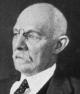
In 1908, a year after this photograph appeared, the modern era of blood transfusion began when Alexis Carrel, MD, sutured a father’s artery to a dying baby’s vein. Cleveland’s George W. Crile, MD (1864–1943), developed a cannula to attach blood vessels and this method, aptly named “direct blood transfusion,” became the procedure of choice for blood transfusion. It was the method of blood replenishment used for decades. In 1900, Karl Landsteiner, MD (1868–1943), discovered the blood groups A, B, and O, and published his findings. Unfortunately, few physicians paid attention to his discovery and for years performed direct blood transfusions hoping there would be no reaction.
Dr. Landsteiner did win the 1930 Nobel Prize in Medicine for his work. By 1915, Dr. Crile and others developed detailed protocols for dealing with surgical shock from blood loss. Dr. Crile also developed a rubber surgical suit for the patient that proved a more effective method for autotransfusion. ■